
2 & 4 Flute End Mill Set, TiN Coated High Speed Steel, 10 ... - little machine shop feeds and speeds
H.F. Poulsen, Three-dimensional X-ray Microscopy — Mapping Polycrystals and their Dynamics (Heidelberg, Germany: Springer, 2004).
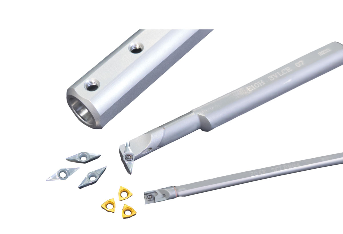
GenSwiss' new mini boring line provides a method of creating ID features that utilizes easy to change ISO style turning inserts and round shank holders.
Available solid carbide insert holders provide enhanced vibration dampening to ensure tool cutting accuracy. Each holder features coolant-thru outlets and shank diameters down to 10mm. Boring holders can be paired with a GenSwiss bushing for easy mounting to static round shank tool holders.
A. Stormvinter, P. Hedström and A. Borgenstam. ” Investigation of Lath and Plate Martensite in a Carbon Steel”, Solid State. Phenom. 172–174 (2011) 61.
S. Morito, Y. Edamatsu, K. Ichinotani, T. Ohba, T. Hayashi, Y. Adachi, T. Furuhara, G. Miyamoto, N. Takayama. “Quantitative analysis of three-dimensional morphology of martensite packets and blocks in iron-carbon-manganese steels”, J. Alloy. Compd., http://dx.doi.org/10.1016/j.jallcom.2012.02.004.
Y. Liu, G. Zhang and W. Li, “A study of martensitic morphology by parallel multiple-layers sectioning”, Acta Metall. Sin. 46 (2010) 930.
J. Kundin, D. Raabe and H. Emmerich. “A phase-field model for incoherent martensitic transformations including plastic accommodation processes in the austenite”, J. Mech. Phys. Solids 59 (2011) 2082.
P. Hedström, U. Lienert, J. Almer and M. Odén. ”Elastic strain evolution and ɛ-martensite formation in individual austenite grains during in situ loading of a metastable stainless steel”, Mater. Lett. 62 (2008) 338.
© 2024 Genevieve Swiss Industries, Inc. | All rights reserved. Privacy Policy | Sitemap Magento development by MageMontreal
Inserts come in 3 geometries: 35 degree rhombic, 80 degree rhombic, and 80 degree trigon. Several chip breaker geometries and coatings are available, ideal for cutting materials such as carbon alloy steels, stainless steels, cast iron, and aluminum alloys.
D.J. Rowenhorst, A. Gupta, C.R. Feng and G. Spanos, “3D Crystallographic and morphological analysis of coarse martensite: Combining EBSD and serial sectioning” Scripta Mater. 55 (2006) 11.
The mechanical properties of high-performance steels are often reliant on the hard martensitic structure. It can either be the sole constituent e.g. in tool steels, or it can be part of a multi-phase structure as e.g. in dual-phase steels. It is well-known that the morphology of martensite changes from lath to plate martensite with increasing carbon content. The transition from lath to plate is however less known and in particular the three-dimensional (3D) aspects in the mixed lath and plate region require more work. Here the current view of the 3D microstructure of martensite in carbon steels is briefly reviewed and complemented by serial sectioning experiments using a focused ion beam scanning electron microscope (FIB-SEM). The large martensite units in the Fe-1.2 mass% C steel investigated here are found to have one dominant growth direction, less transverse growth and very limited thickening. There is also evident transformation twinning parallel to the transverse direction. It is concluded that more 3D analysis is required to understand the 3D microstructure of martensite in the mixed lath and plate region and to verify the recently proposed 3D phase field models of martensite in steels.
S. Morito, Y. Adachi and T. Ohba. “Morphology and Crystallography of Sub-Blocks in Ultra-Low Carbon Lath Martensite Steel” Mater. Trans. 50 (2009) 1919.
Hedström, P. et al. (2012). On the Three-Dimensional Microstructure of Martensite in Carbon Steels. In: De Graef, M., Poulsen, H.F., Lewis, A., Simmons, J., Spanos, G. (eds) Proceedings of the 1st International Conference on 3D Materials Science. Springer, Cham. https://doi.org/10.1007/978-3-319-48762-5_3
J.R. Kremer, D.N. Mastronarde and J.R. McIntosh. “Computer visualization of three-dimensional image data using IMOD”, J. Struct. Biol. 116 (1996) 71.
H.K. Yeddu, A. Borgenstam, P. Hedström and J. Ågren. ”A phase-field study of the physical concepts of martensitic transformations in steels” Mater. Sci. Eng. A 538 (2012) 173.
H.K. Yeddu, A. Malik, J. Ågren, G. Amberg, A. Borgenstam. ”Three-dimensional phase-field modeling of martensitic microstructure evolution in steels”, Acta Mater. 60 (2012) 1538.
A. Stormvinter, P. Hedström and A. Borgenstam. ”Automatic phase and orientation mapping combined with classical transmission electron microscopy to study the development of martensite in high-carbon low alloy steels”, Submitted 2012.
S. Zaefferer and S.I. Wright. “Three-dimensional orientation microscopy by serial sectioning and EBSD-based orientation mapping in a FIB-SEM”, Electron backscatter diffraction in materials science, ed. A.J. Schwartz, M. Kumar, B.L. Adams and D.P. Field (New York, NY: Springer, 2009) 109–122.
JavaScript seems to be disabled in your browser. For the best experience on our site, be sure to turn on Javascript in your browser.


 0086-813-8127573