27315: CS700H 24 V Power Supply Kit - 27315

Oxford Academic is home to a wide variety of products. The institutional subscription may not cover the content that you are trying to access. If you believe you should have access to that content, please contact your librarian.
Results: Complications occurred in six of the 60 patients (10%) who underwent bilateral otoplasty. A discrete recurrence occurred in the superior pole in six patients (10%) and was unilateral in all cases. Follow-up ranged from three months to 10 years. There were no major recurrences in this series. The satisfaction rate was 98.3%. The antihelix looked smooth, without marks or irregularities; the helix seemed straight and well placed. The posterior scar was inconspicuous.
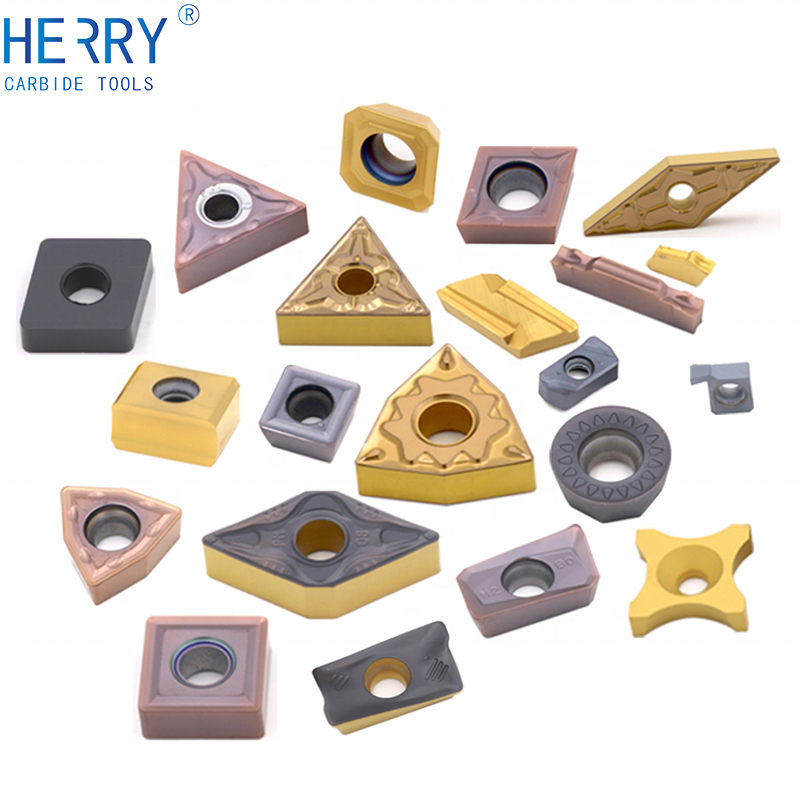
Improving the external machining processes is essential for staying ahead of the competition. If you're looking for external machining tools that offer consistent and accurate results, you've come to the right place. At Kennametal, we design and manufacture industry-leading turning systems that leverage the most up-to-date technology. With consistent and accurate results our external tooling combat wear and tear while minimizing costly downtime. If we do not already have the positive and negative clamping styles and tool holders you need for your applications, our design engineers can help you customize the right Kennametal turning tools for all your business operations.
Typically, access is provided across an institutional network to a range of IP addresses. This authentication occurs automatically, and it is not possible to sign out of an IP authenticated account.
Access to content on Oxford Academic is often provided through institutional subscriptions and purchases. If you are a member of an institution with an active account, you may be able to access content in one of the following ways:
Conclusions: The present technique introduces a new concept in otoplasty that avoids handling the antihelix cartilage. A single incision of the antihelix breaks the cartilage spring memory completely and allows rebuilding of the antihelical fold without resorting to techniques such as rasping, drilling, excision, or complete incision, all of which are time-consuming and may result in irregularities. Sutures are placed to create the new antihelix in the desired shape easily and without tension. The helix and earlobe are repositioned with a specific posterior skin resection and with the tail of the helix’s replacement.
Many societies offer single sign-on between the society website and Oxford Academic. If you see ‘Sign in through society site’ in the sign in pane within a journal:
Background: For over a century, various otoplasty techniques for correction of the prominent ear have been attempted. Nevertheless, cartilage memory strength, mainly in the thick cartilage (and consequently the recurrence of the prominence), still remains a problem. An additional difficulty relates to the antihelix irregularities caused by attempts to weaken the cartilage spring, which are time-consuming and in some cases lead to an unnatural-looking result.
Choose this option to get remote access when outside your institution. Shibboleth/Open Athens technology is used to provide single sign-on between your institution’s website and Oxford Academic.
Objective: The technique reported in this article represents a new approach to otoplasty that deconstructs the auricular pavilion with a single full incision in the antihelix. This approach makes it easier to rebuild the antihelical fold with dissolving sutures and allows repositioning of the helix and earlobe with a posterior bielliptical incision and a narrow bridge skin resection.
Augusto Sette Câmara Valente, Separating the Helix From the Antihelix: A New Concept in Prominent Ear Correction, Aesthetic Surgery Journal, Volume 30, Issue 2, March 2010, Pages 139–153, https://doi.org/10.1177/1090820X10369689
Oxford University Press is a department of the University of Oxford. It furthers the University's objective of excellence in research, scholarship, and education by publishing worldwide
For librarians and administrators, your personal account also provides access to institutional account management. Here you will find options to view and activate subscriptions, manage institutional settings and access options, access usage statistics, and more.
Methods: Between 1999 and 2009, the author performed otoplasty for correction of the prominent ear in 60 patients, including 32 men (53.3%) and 28 women (46.7%). A single incision was made in the superior and lateral borders of the antihelix from the triangular fossa down to the tail of the helix, to completely separate the antihelix from the helix. Three postauricular sutures were applied with nonpermanent side-to-side mattress sutures, with a fourth suture placed to reposition the tail of the helix. A posterior bielliptical incision with a narrow bridge skin resection was performed.
. Please enter the desired qty for the material(s) you want to include in your promotion or Proceed Without Promotion and only your base materials will be added to the cart.
If your institution is not listed or you cannot sign in to your institution’s website, please contact your librarian or administrator.


 0086-813-8127573